ALS is a progressive neurodegenerative disease characterized by cumulative dysfunction and eventual death of motor neurons, which is why it is sometimes referred to as Motor Neuron Disease (MND). However, let’s take a step back and start with a brief summary of neurons and their role in the nervous system before diving into the biological basis of the disease.
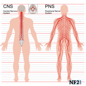
The human nervous system is divided into the central nervous system (CNS), which is composed of the brain and spinal cord, and the peripheral nervous system (PNS), which includes any nerves and nervous tissue not included in the CNS. The CNS and PNS are connected together by strings of neurons.
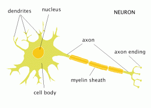
Tissue in the nervous system is primarily composed of neurons and support cells (or glial cells). Neurons have three parts: The cell body, which contains the nucleus, multiple dendrites, which gather signals from surrounding neurons and bring them to the cell body, and a single axon, which transmits a signal from the cell body to the surrounding nervous tissue.
There are three primary types of neuron:
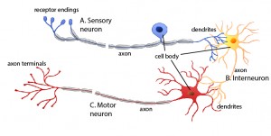
1. Sensory Neurons – receive input of environmental information from sensory organs (eyes, ears, skin, nose, mouth), and carry these messages to the brain.
2. Interneurons – connect neurons together by transmitting messages (usually connect the sensory neurons to motor neurons).
3. Motor Neurons – gather information (usually from interneurons, which have passed along information gathered by sensory neurons), and transmit impulses to bodily muscles, resulting in movement.

(Figure 3) A depiction of the spatial relationships of the three types of neurons. The interneuron acts as the bridge that conducts signals from sensory neurons to motor neurons.
There are two different subtypes of motor neurons:
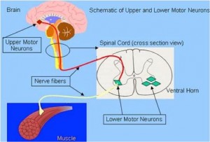
1. Upper Motor Neurons (UMNs) – originate in the motor cortex of the brain, and carry electrical impulses to the lower motor neurons.
2. Lower Motor Neurons (LMNs) – originate in the spinal cord, gather impulses from UMNs and transmit them to skeletal muscles.

(Figure 4) A diagram depicting the connection between upper and lower motor neurons. UMNs are found in the motor cortex, while LMNs are found in the brain stem and spinal cord.

(Figure 5) Motor cortex location in the brain. This is the site where upper motor neurons are found.
While many different neurological disorders affect just one of these motor neuron subtypes, ALS, unfortunately, affects both. As motor neurons degenerate, their ability to formulate and transmit impulses declines, which leads to loss of motor control. Early symptoms are consistent with small neurological deficits in certain muscle groups: frequent tripping, localized muscle weakness, nasal or slurred speech, and muscular twitching all result from the early decline of neurological tissue. As the disease progresses and more motor neurons die, the symptoms are exacerbated to the point that individuals often become wheelchair-bound, and lose all motor control. Most people succumb to respiratory failure 3-5 years after diagnosis due to degeneration of motor neurons that innervate to the diaphragm and intercostal muscles. It is important to emphasize that muscular atrophy occurs as a result of nervous tissue degeneration and that the muscles, themselves, are disease free. That is, the disorder lies in the motor neurons, not the muscles that they innervate.
Figures were derived from the following links
Figure 1: http://www.nf2is.org/nerves.php
Figure 2: http://webspace.ship.edu/cgboer/theneuron.html
Figure 3: http://cnx.org/contents/a4ca89c4-ab19-492f-a910-8f0f7867999f@6/Nervous-System
Figure 4: https://www.studyblue.com/notes/note/n/l3-degenerative-disease-and-dementia/deck/14274590
Figure 5: http://hirnforschung.kyb.mpg.de/en/brain-research.html
References:
- Beghi E, Mennini T; Italian Network for the Study of Motor Neuron Disease. 2004. Basic and clinical research on amyotrophic lateral sclerosis and other motor neuron disorders in Italy: recent findings and achievements from a network of laboratories. Neurol Sci 25 Suppl 2:S41-60.
- Gagliardi S, Emanuela C, Davin A, Guareschi S, Abel K, Alvisi E, Laforenza U, Ghidoni R, Cashman JR, Ceroni M, Cereda C. 2010. SOD1 mRNA expression in sporadic amyotrophic lateral sclerosis. Neurobiology of Disease 39(2):198-203.
- Manfredi G, Zuoshang X, 2005. Mitochondrial dysfunction and its role in motor neuron degeneration in ALS. Mitochondrion 2005(5):77-78.
- Mühling T, Duda J, Weishaupt JH, Ludolph AC, Liss B. 2014. Elevated mRNA-levels of distinct mitochondrial and plasma membrane Ca2+ transporters in individual hypoglossal motor neurons of endstage SOD1 transgenic mice. Frontiers in Cellular Neuroscience 353(8):1-14.
- Van Zundert B, Izaurieta P, Fritz E, Alvarez FJ. 2012. Early pathogenesis in the adult-onset neurodegenerative disease amyotrophic lateral sclerosis. J Cell Biochem 113(11):3301-12.

